Staging
Staging Investigations
Liver tumours may be staged by radiological investigations including ultrasound (US) scanning, Spiral Computed Tomography (CT) scanning Magnetic Resonance Imaging (MRI) and Positron Emission Tomography (PET) scanning.
The evidence would suggest that multiple forms of imaging are often required to identify and characterise different types of liver lesion. Ultrasound is relatively cheap and easy to perform although the results are operator dependent.
CT Scanning
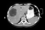
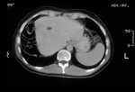
CT scanning is the most commonly used technique and with the advent of contrast enhanced spiral CT scanning definition of liver lesions has improved enormously.
Magnetic Resonance Imaging (MRI)
MRI with Teslascan has improved the sensitivity and specificity of MRI imaging hugely and this technique is highly sensitive. Recently our Unit has begun to evaluate Prima Vist.
 |
 |
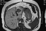
| Figure 3 is an MRI showing lesion in segment 3 of the left hemi-liver. |

Figure 3 - click image to enlarge |
Laparoscopy
Laparoscopy, carried out as a day procedure prior to formal surgical exploration or actually carried out on the day of main surgery, is useful for spotting small nodules of disseminated disease not evident on ultrasound, CT or MRI scanning. When combined with laparoscopic ultrasound the technique is even more accurate. Widespread adoption of this technique has reduced the number of 'open and shut' cases in one series from 13% to 7%.
Operative ultrasound
Operative ultrasound carried out at the time of surgery is the most sensitive investigation available provided that the operating surgeon has a high quality machine with a special liver probe.

